Adolescents aged between 7 and 20 years, particularly those with a Haller Index greater than 3.25 or with clinical manifestations of cardiopulmonary compression
Pectus excavatum is the most common congenital deformity of the chest wall, predominantly affecting male adolescents. It is characterized by posterior displacement of the sternum, which compresses thoracic organs and results in decreased cardiopulmonary capacity. Traditional treatment mainly involves the minimally invasive Nuss procedure. While it provides significant aesthetic and functional improvements, challenges remain for patients with severe or asymmetric deformities. The Pediatric Surgery Department at Tri-Service General Hospital has developed an innovative approach that combines 3D thoracic printing and the sternal elevation technique to address these issues more safely and effectively. With 3D printing, we generate patient-specific thoracic models from CT scans, enabling preoperative simulation and the design of custom-shaped correction bars. The sternal elevation technique allows intraoperative lifting of the sternum to enlarge the surgical field and avoid injury to vital organs. This strategy has been successfully applied in 40 patients, all of whom experienced significant improvements without major complications. This innovation exemplifies the integration of precision medicine, surgical safety, and patient-centered care.
The innovative surgical treatment for pectus excavatum encompasses a fully standardized workflow covering the entire continuum from precise preoperative imaging and modeling to long-term postoperative rehabilitation and follow-up.
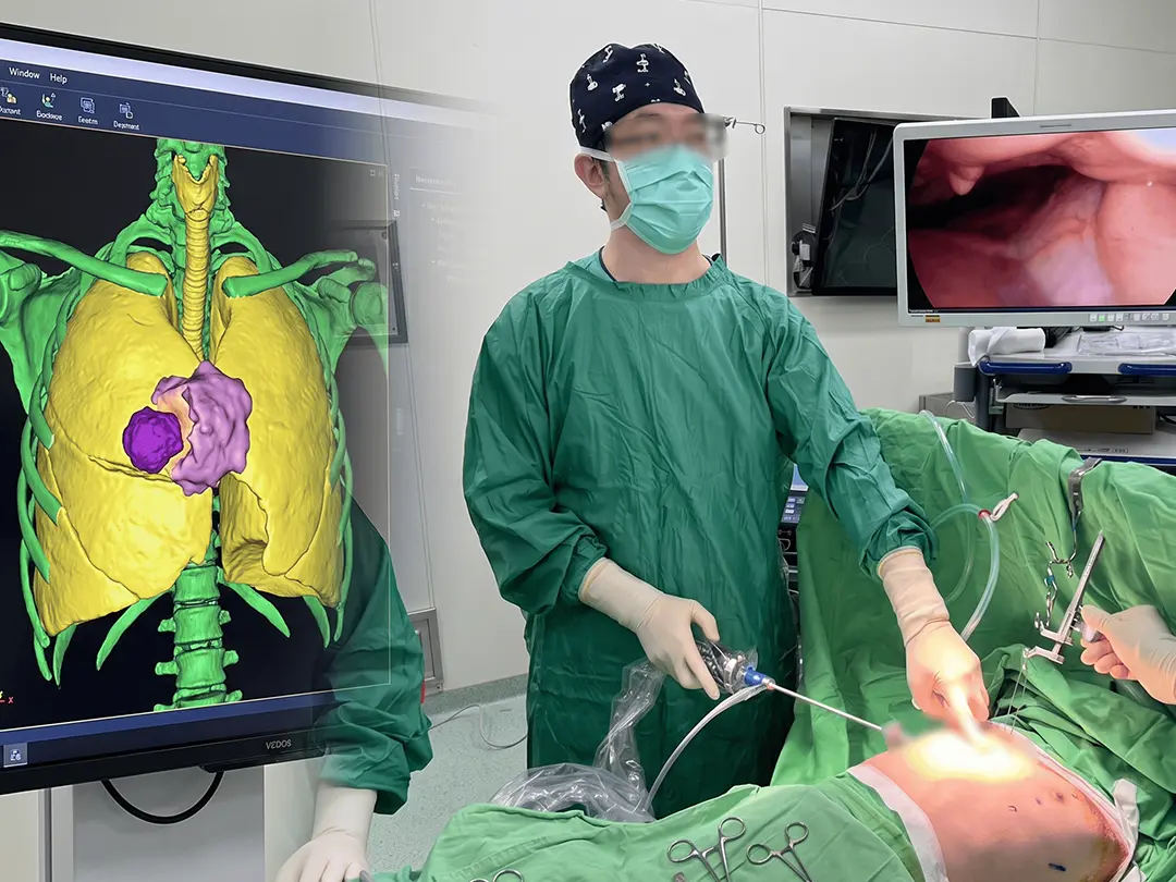
Preoperative high-resolution chest computed tomography (CT) with construction of 3D-printed simulation models.
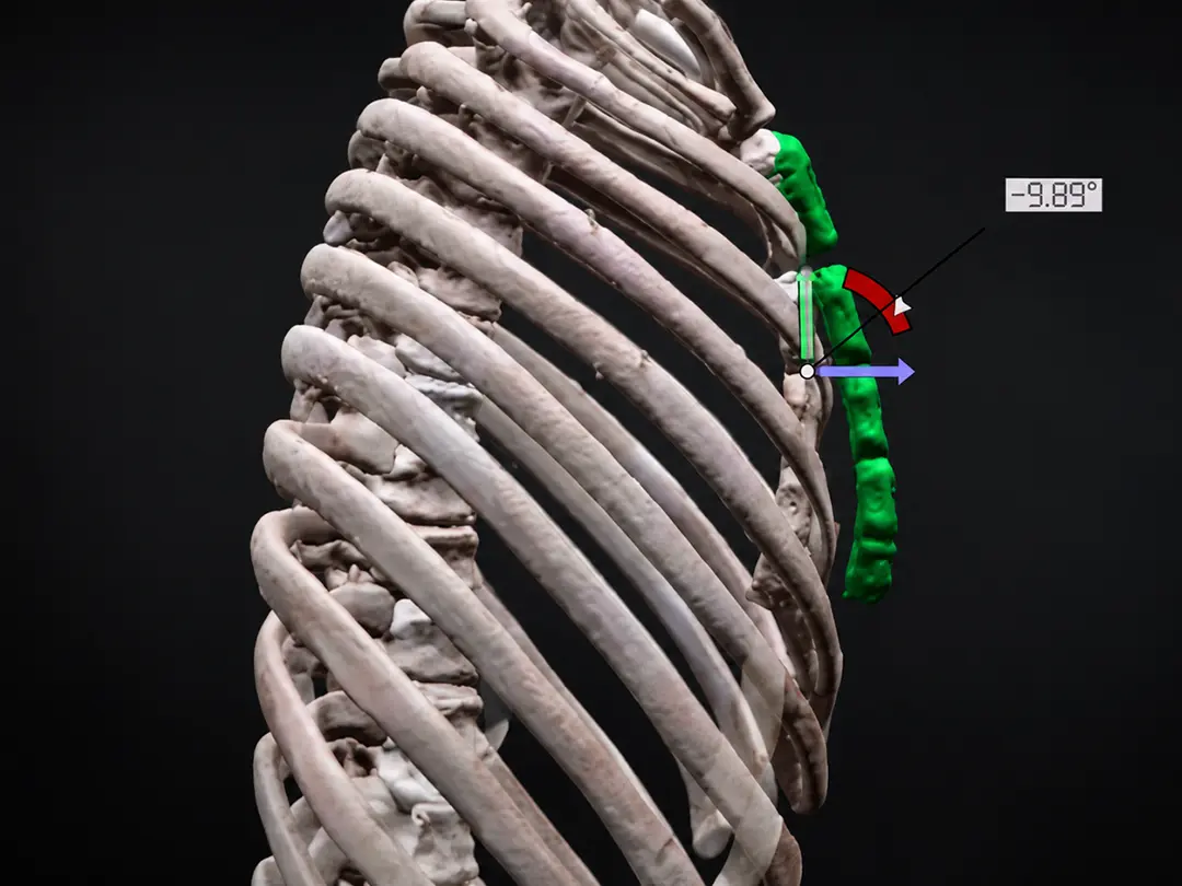
Design and pre-bending of customized corrective steel bars to conform to individual chest wall anatomy, thereby enhancing fixation stability.
Intraoperative application of the sternal elevation (sternal suspension) technique to initially lift the depressed area, establishing a safe operative field and preventing injury to the heart, lungs, and major vessels.
Performance of the Nuss procedure under thoracoscopic assistance, reducing invasiveness and improving intraoperative visualization.
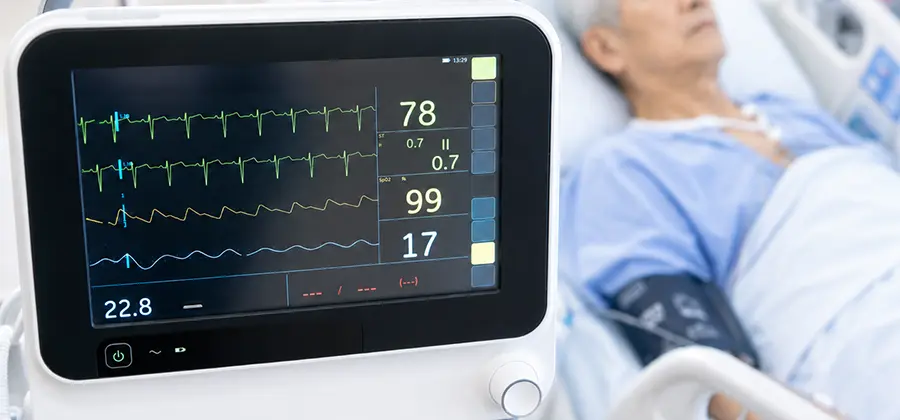
Postoperative pain management, intensive care unit monitoring, and structured rehabilitation planning to ensure optimal recovery outcomes.
From July 2022 to February 2025, a total of 40 patients were treated, all of whom were managed in accordance with a standardized protocol of “prediction–simulation–design–execution–follow-up.”
The mean preoperative Haller Index was 3.88, which decreased to 2.56 postoperatively.
The average length of hospital stay was 4.7 days, representing a reduction of more than 30% compared with the traditional approach (6–8 days).
The mean operative time was shortened to 86.6 minutes, with intraoperative blood loss of less than 10 mL.
The overall intraoperative complication rate was 2.5% (1/40), with no cases of cardiac, pulmonary, or major vascular injury.
No common complications were observed, such as pneumothorax, hemothorax, pleural effusion, or displacement of the corrective bar.
Follow-up at 6 to 12 months revealed no adverse events, indicating that this treatment protocol demonstrates long-term stability.
Patients and their families expressed a high level of satisfaction with the preoperative model-based explanations, intraoperative procedural explanations, postoperative pain management, and rehabilitation planning.
Most families reported that the surgical outcomes exceeded expectations and that the 3D models facilitated a better understanding of the surgical approach, clearly reflecting the effectiveness of clinical communication and patient-centered care.
The overall satisfaction score reached 4.8 out of 5.
The novel design model of 3D printing–assisted customized corrective bars for pectus excavatum received the National Innovation Award in 2017.
Related research findings were published in the journal of the American Association for Thoracic Surgery (The Annals of Thoracic Surgery) and were recognized by the international academic community.
In the future, this approach will be expanded to other indications for chest wall reconstruction, promoting the widespread adoption of personalized medicine applications.
A domestically pioneering and innovative surgical strategy for pectus excavatum correction, integrating 3D chest wall printing and sternal elevation techniques, provides a safer and more effective surgical approach for patients with difficult and complex forms of pectus excavatum.
3D-printed chest wall models are used to simulate postoperative thoracic appearance and to preoperatively plan and custom-measure appropriate corrective bars.
The sternal elevation technique significantly enhances surgical safety and provides a superior operative field.
This approach reduces anxiety and feelings of insecurity among patients and their families regarding pectus excavatum surgery, thereby improving patient confidence and satisfaction.
Excellent outcomes are achieved in terms of chest wall appearance improvement, surgical safety, and reduction of postoperative complications.