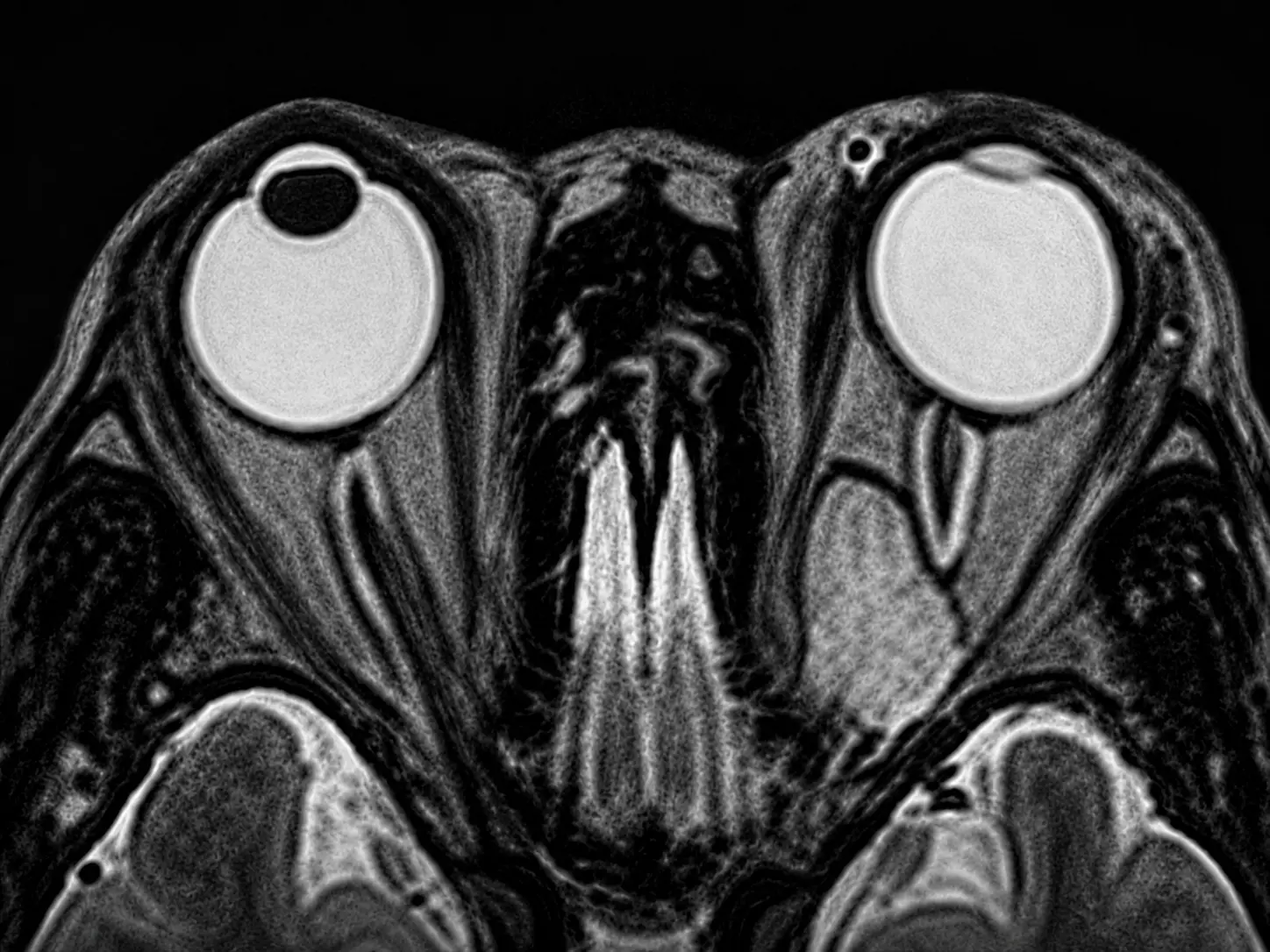
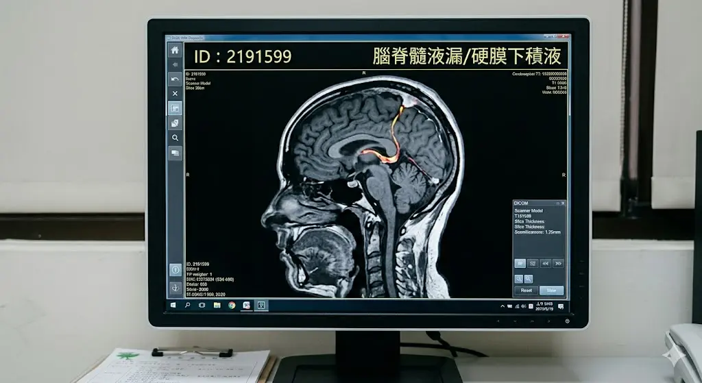
.Benign or malignant brain tumors.
.Meningioma, acoustic neuroma, or pituitary tumors.
.Brain metastases or deep brain lesions.
.AVMs or vascular lesions.
.Trigeminal neuralgia.
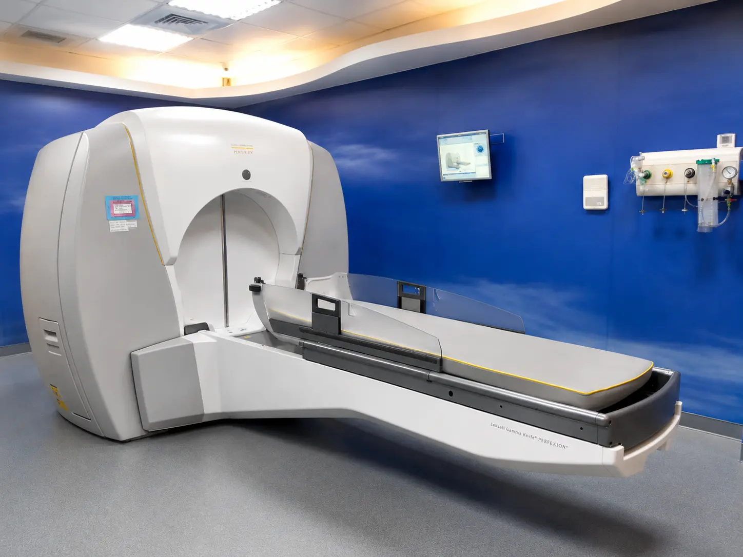
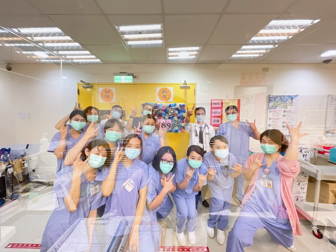
When our hospital was founded in 1999, we installed the most advanced Gamma Knife radiosurgery machine, established the Gamma Knife Center, and recruited brain radiosurgery talents. The current members of the Gama Knife Center include a chief neurosurgeon, five full-time and part-time attending physicians, a radiation physicist, a case manager and two nurses. The above-mentioned members include interdisciplinary personnel from neurosurgery, radiation oncology, and diagnostic radiology, forming a professional and all-round treatment team that adheres to a "patient-centered" service and promotes whole-person, full-responsibility and long-term follow-up care. , create a safe, comfortable and warm medical environment to serve patients with brain tumors and vascular lesions, and rigorously track the patient's condition over the long term.
. Precise focusing of 192 Cobalt-60 beams on the lesion.
. Stereotactic localization combined with 3D image-based planning.
. Radiation targeting error of less than 0.5 mm.
. Treatment without craniotomy or general anesthesia.
. Multidisciplinary team comprising neurosurgery, radiation oncology, and radiology specialists.
. Long-term treatment efficacy monitoring via an electronic database.
. AI-assisted image analysis to support treatment decision-making.
Our center treats an average of about 180-200 patients every year. Treatment of intracerebral lesions includes: brain arteriovenous malformation (8.1%); brain arteriovenous fistula (3.3%); cavernous malformations (3.4%); meningioma (17.5%); acoustic neuroma (15.6%); Pituitary gland tumors (5.8%); craniopharyngioma (1.3%); metastatic brain tumors (18.6%); trigeminal neuralgia (14.9%) and other tumors (11.5%); from the hospital's establishment in 1999 to recently By the end of August 2024, the Gamma Knife Center had treated a total of 2,599 patients, providing safe and effective treatment for domestic deep brain tumor patients.
.Control rate for benign brain tumors: 87%–98%.
.Complete resolution of AVMs in 77 cases.
.Overall AVM resolution rate: 65.3%.
.Improvement rate for trigeminal neuralgia: 82%.
.Medication discontinuation rate for trigeminal neuralgia: 55%.
.Cumulative total of 2,243 treatment sessions.
.Average of approximately 225 treatment sessions per year.
.Radiation margin of error < 0.5 mm.
Pre-treatment safety checks:
Pre-treatment "Time-out" verification: The attending physician, technician, or nursing staff must perform a double-check to verify the following: the consent form is correctly and fully completed, the patient's identity is correct, the procedure/treatment type is correct, the treatment site is correct, and the equipment is verified.
Frame location check: Confirm that the margin of error is ≤1 mm.
Spot check: Perform a spot check immediately prior to treatment to confirm the accuracy of the head frame's position.
Clearance test: Measure the head frame and pin lengths, and conduct a clearance test before treatment to prevent the head frame from colliding with the machine's internal components.
Mask location check: Before each treatment session, a Cone Beam CT scan is performed to assess positioning errors. Following Cone Beam CT localization, the system automatically adjusts the 6D coordinate axes (X, Θx, Y, Θy, Z, Θz) before proceeding with the patient's treatment.
.Non-invasive treatment (no craniotomy required), reducing physical and mental burden.
.Rapid return home and to work following treatment.
.Case management and long-term follow-up provided.
.Regular MRI monitoring to track treatment efficacy.
.Safe and comfortable medical environment.
.Integrated care provided by a multidisciplinary team.
.Published 14 SCI-indexed papers over the past five years.
.Research cited by international Gamma Knife centers.
.Participated in conferences of the Asian Gamma Knife Society.
.Invited to speak at the Russian Medical Association.
.Invited to speak at a neurosurgery conference in Vietnam.
.Collaborated on medical personnel training with the Marshall Islands.
.Presented internationally on the implementation of AI-based image analysis.
.Provides minimally invasive treatment for deep-seated brain lesions.
.Reduces the risks associated with craniotomy and anesthesia.
.Shortens hospital stays, allowing for a quick return home after treatment.
.Preserves normal brain tissue and neurological function.
.Enhances treatment quality through long-term follow-up.
.Establishes a multidisciplinary model for precision treatment.
.Builds a database to support AI applications and research.