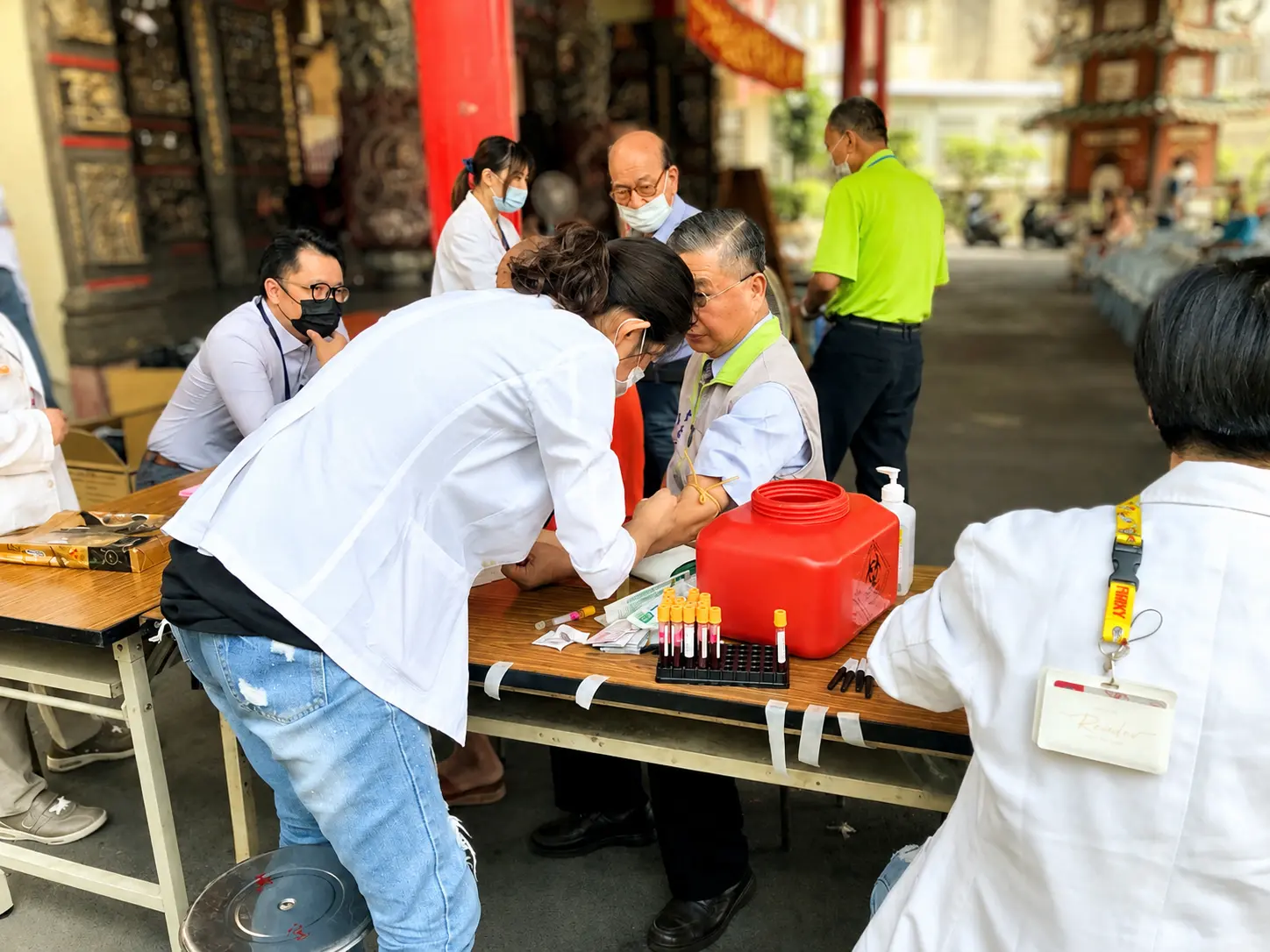
Men over 50 take screening including PSA testing and digital rectal examination. If abnormalities are found, PHI, mpMRI, or MRI-ultrasound fusion biopsy may help improve diagnostic accuracy.
PSA specificity is relatively low and can be influenced by factors such as benign prostatic hyperplasia (BPH) and urinary tract infections. Relying solely on PSA levels for biopsy decisions may lead to overdiagnosis and overtreatment. The Prostate Health Index (PHI), calculated using PSA, free PSA, and p2PSA through a mathematical algorithm, offers greater accuracy compared to PSA alone.
Traditional transrectal ultrasound (TRUS)-guided prostate biopsy cannot identify tumor locations on ultrasound images, relying instead on systematic, random sampling. Accurately targeting the tumor with biopsy needles is akin to finding a needle in a haystack. Multiparametric MRI (mpMRI) can assess prostate cancer risk, localize tumor regions, and aid in staging the cancer.
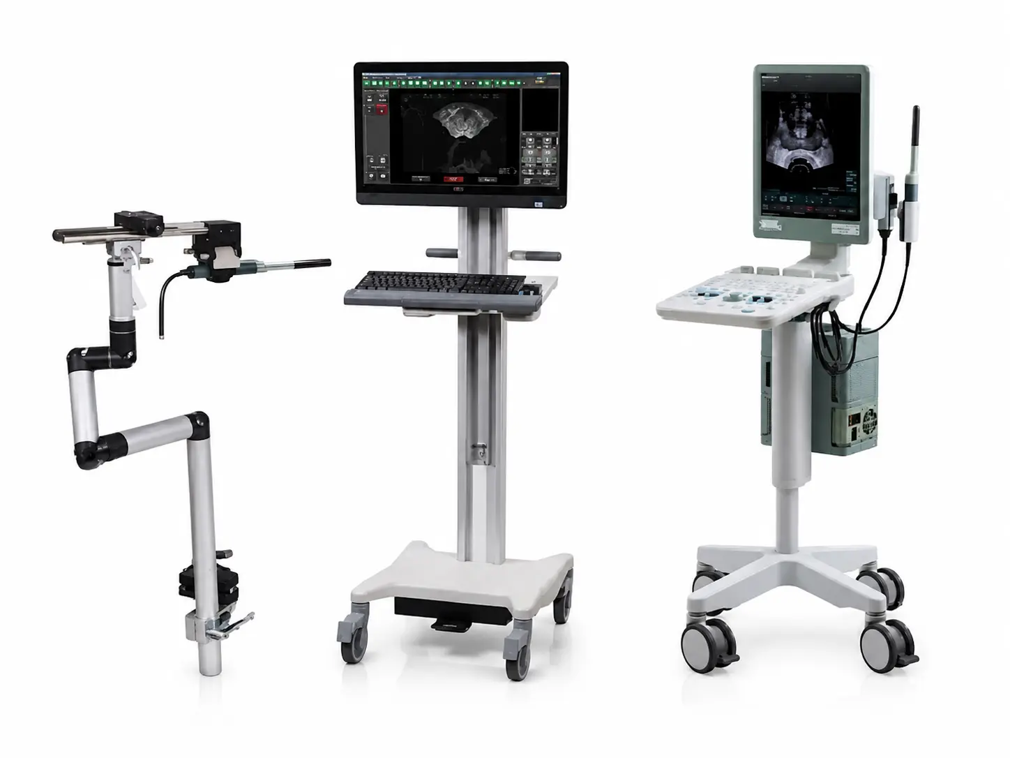
MRI-ultrasound fusion imaging combines reconstructed 3D MRI images of the prostate and tumor with real-time ultrasound images using computer software. This allows clear visualization of the tumor on ultrasound. Targeted biopsy can then be performed, significantly improving biopsy accuracy.
High-quality prostate mpMRI and radiologists specializing in mpMRI interpretation can clearly differentiate between benign prostatic hyperplasia and prostate cancer, accurately marking the tumor's location and extent before biopsy. Dr. Lin Wei-Ching, Director of Radiology, has undergone advanced training in prostate mpMRI in the United States and South Korea. Over the years, she has gained extensive experience by repeatedly correlating mpMRI findings with tumor pathology reports, achieving increasingly precise interpretations.
Urologists analyze various clinical data, such as patient age, PSA, digital rectal examination (DRE), PHI, and mpMRI, to assess the likelihood of prostate cancer and determine the necessity of a biopsy, thereby avoiding unnecessary procedures.
Urologists proficiently operate image fusion systems to accurately integrate mpMRI with ultrasound images. They are skilled in transperineal biopsy techniques and understand the concept of saturation and targeted biopsy. Dr. Hsieh Po-Fan, a urologist, has undergone advanced training in MRI-ultrasound image fusion-guided prostate biopsy in the United States and Japan. Through regular multidisciplinary meetings with pathology, radiology, and urology teams, as well as consultations with international experts, they identify and address initial errors such as patient positioning, level alignment, and biopsy needle details. With accumulated experience, their techniques have become increasingly refined, enabling precise tumor targeting during biopsy under local anesthesia, minimizing the risks associated with general anesthesia.
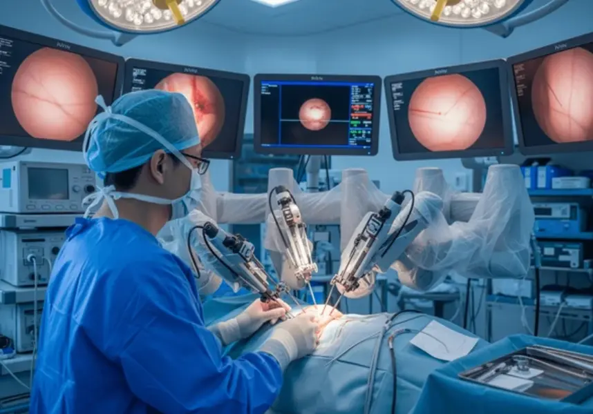
With accurate tumor localization and clinical risk assessment, some early-stage prostate cancer patients are identified as suitable candidates for focal HIFU (High-Intensity Focused Ultrasound) treatment, significantly reducing the risks of postoperative urinary incontinence and sexual dysfunction. They are well-versed in HIFU equipment operation and energy monitoring.
1. Number of Patients Undergoing MRI and Ultrasound Fusion Biopsy
2022: 112 patients
2023: 148 patients
January to September 2024: 98 patients
2. HIFU Procedures
2022: 9 patients
2023: 10 patients
January to September 2024: 13 patients
Cancer detection rate using MRI-ultrasound fusion biopsy: 59%
Cancer detection rates for PI-RADS scores 3 to 5: 28% / 45% / 77%
Transperineal biopsy
AUR: 18.5%
UTI: 1.1%
Sepsis: 0
Gross hematuria: 11.9%
Hemospermia: 0%
Pain on biopsy site: 6.5%
Perineal hematoma: 3.2%
With the collective efforts of the entire medical team, all patients receive regular follow-up care after treatment. This enables us to precisely monitor each patient's real-time condition, providing them with the safest and most reassuring treatment environment.
Published findings on the pathological characteristics of tumors undetectable by mpMRI:
The amount of prostate stroma, severe inflammation, small tumor volume, and growth patterns of the prostate gland affect the detection rate of tumors by mpMRI.
Collaborative publication with other medical centers on PHI density:
PHI density demonstrates a higher detection rate for prostate cancer.
Asia's first study combining mpMRI and PHI for prostate cancer detection:
In our prospective study, the sensitivity, specificity, positive predictive value (PPV), and negative predictive value (NPV) of mpMRI with PI-RADS ≥3 for predicting clinically significant prostate cancer were 100%, 44.9%, 35.8%, and 100%, respectively. For PHI >30, the sensitivity, specificity, PPV, and NPV were 91.7%, 43.6%, 33.3%, and 94.4%, respectively. In ROC curve analysis, combining mpMRI and PHI yielded a higher AUC for predicting clinically significant prostate cancer compared to mpMRI or PHI alone (AUC 0.873 vs. 0.83, p = 0.035; 0.873 vs. 0.735, p = 0.002).
The world's first study combining mpMRI and PHI to predict tumor size:
Analyzing preoperative mpMRI, PHI, and postoperative pathology reports from patients undergoing robotic-assisted radical prostatectomy at our hospital, we found that mpMRI often underestimated tumor extent, especially in lesions with PI-RADS 4 or 5. For PI-RADS 3 tumors, the maximum difference between imaging and pathological lesion diameter was 2 mm. Extending the tumor boundary on mpMRI by 1 mm was sufficient to encompass the entire tumor. For PI-RADS 4 or 5 tumors, with PHI <30, 30-60, and >60, the maximum differences were 12, 16, and 22 mm, respectively. Thus, tumor boundaries on mpMRI need to be extended by 6, 8, and 11 mm to fully cover the tumor extent. In summary, combining mpMRI and PHI allows for a more precise delineation of tumor boundaries, enabling better surgical planning in future procedures.
Weekly Multidisciplinary Imaging Conference (Radiology and Urology)
Patients undergo mpMRI before biopsy. Dr. Lin Wei-Ching, Director of Abdominal Imaging Diagnostics at the Department of Radiology and Consultant at the Center for Innovation in Intelligent Medical Technology, personally interprets the images. Each lesion is assessed with a PI-RADS score, and lesion boundaries are delineated to evaluate prostate cancer risk, locate tumor regions, and assist in staging cancer.
Biweekly Multidisciplinary Pathology Conference (Pathology, Radiology, and Urology)
After patients undergo MRI-ultrasound fusion-guided prostate biopsy, their pathology reports are reviewed to validate mpMRI findings. If discrepancies arise between pathology and imaging reports, the team thoroughly discusses potential issues such as missed imaging findings, fusion errors, or biopsy inaccuracies. Each case discussion drives the team's collective growth and experience.
Biweekly Multidisciplinary conference(Urology, Pathology, Radiology, Hematology-Oncology, and Radiation Oncology)
Urologists lead the team, coordinating efforts across specialties. The team references the NCCN (National Comprehensive Cancer Network) and EAU (European Association of Urology) guidelines while tailoring treatment protocols to local circumstances in Taiwan. This ensures personalized, precision medicine for every patient.
Reducing Infections
Transperineal biopsy significantly reduces the risk of sepsis compared to traditional transrectal biopsy. This approach minimizes the use of prophylactic antibiotics, thereby reducing bacterial resistance in the environment.
Technical Knowledge Transfer
Radiologists interpreting mpMRI and urologists performing image-fusion biopsies receive direct training and supervision from Dr. Lin Wei-Ching and Dr. Hsieh Po-Fan. New physicians are carefully guided and monitored throughout the learning process until they are proficient enough to operate independently.